Neuro Anatomy & Neuro Physiology
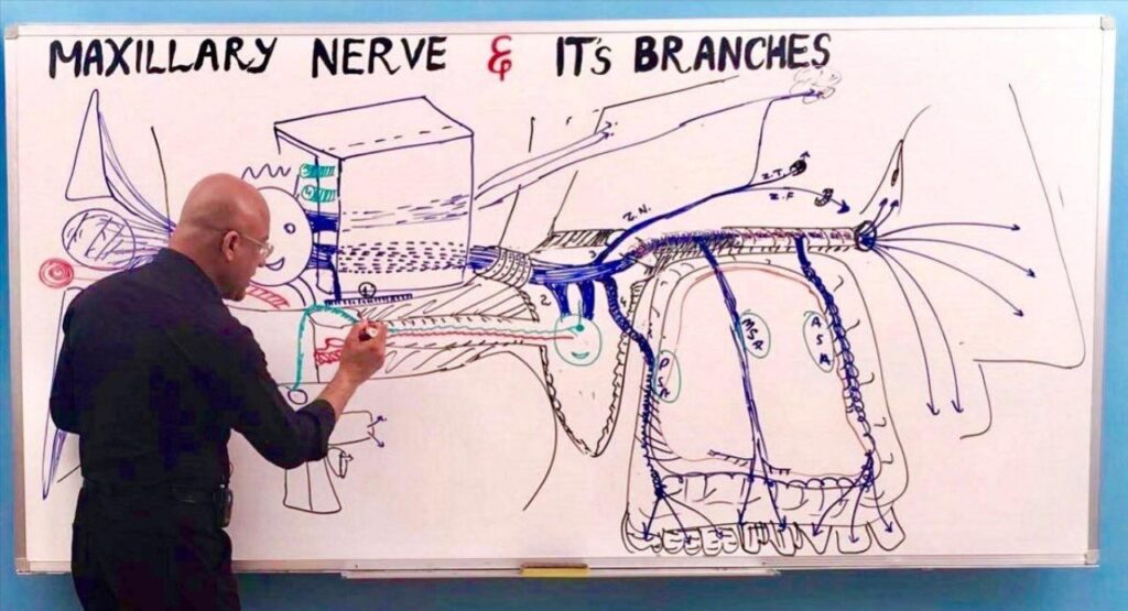
Dr. Najeeb’s Neuro Anatomy & Neuro Physiology course is a series of 138 video lecture parts that teaches the nervous system from the ground up. It covers development, meninges and CSF, the spinal cord and its tracts, the brainstem, cranial nerves, the eye and ear pathways, the basal ganglia and the autonomic nervous system. Every structure and pathway is drawn live on a whiteboard, so you see how it fits together instead of memorising it. Handwritten notes are available for every topic in the course.
| Course | Neuro Anatomy & Neuro Physiology |
|---|---|
| Instructor | Dr. Najeeb, over 40 years of teaching experience |
| Content | 138 lecture parts across six modules |
| Teaching style | Hand-drawn, step-by-step whiteboard lectures |
| Level | Beginner-friendly; no prior medical knowledge needed |
| Access | Lifetime access with the premium plan, including future lectures |
| Notes | Handwritten notes for every topic, sold separately (Notes add-on or Combo Plan) |
| Free preview | Available through a free “Try Us Free” account |
| Suitable for | Medical, dental, nursing, pharmacy and physiotherapy students; USMLE Step 1, PLAB, COMLEX and NEET PG preparation |
What you will learn
The course is built so each module prepares you for the next. By the end you should be able to:
- Explain how the nervous system develops, and how the meninges, CSF and blood supply protect and nourish the brain.
- Trace the ascending and descending spinal pathways and predict the deficits after a spinal cord lesion.
- Tell upper motor neuron lesions from lower motor neuron lesions.
- Describe the nuclei, tracts and functions of the medulla, pons, midbrain, cerebellum, thalamus and hypothalamus.
- Follow each cranial nerve from its nucleus to its target, and link a lesion to the signs it produces.
- Explain how hearing, balance, vision and eye movements work.
- Describe the basal ganglia, cerebral cortex, speech areas and the autonomic nervous system, including catecholamine synthesis and release.
Clinical topics covered
The lectures keep returning to the clinic. These topics have their own parts in the series:
- Spinal cord lesions and Brown-Séquard syndrome
- Upper and lower motor neuron lesions
- Oculomotor, trochlear, abducens and facial nerve lesions
- Horner’s syndrome and the oculosympathetic pathway
- Cavernous sinus anatomy and cavernous sinus syndrome
- Bulbar and pseudobulbar palsy
- Horizontal conjugate eye movements and the vestibulo-ocular reflex
- Epilepsy
Course curriculum
| Module | Topics (number of lecture parts) | Parts |
|---|---|---|
| 1. Foundations and protection of the CNS | Introduction to Neuro-Anatomy (2); Development of the Nervous System (2); Meninges (2); CSF and Ventricular System (5); Blood Supply to the Brain (2); Blood-Brain Barrier and Circumventricular Organs (2); Histology of the Nervous System (1) | 16 |
| 2. Spinal cord, tracts and motor neurons | Spinal Cord and its Lesions (3); Brown-Séquard Syndrome (1); Ascending Tracts (4); Motor System and Descending Tracts (3); Upper and Lower Motor Neurons and their Lesions (3); Typical Spinal Nerve (2) | 16 |
| 3. Brainstem, cerebellum and diencephalon | Brain Stem Basic Concepts (3); Medulla (4); Pons (2); Mid-Brain (2); Cerebellum (3); Diencephalon (2); Thalamus (3); Hypothalamus (4) | 23 |
| 4. Cranial nerves and head ganglia | Cranial Nerves Basic Concepts (3); Oculomotor Nerve and Clinical Correlates (6); Trochlear Nerve (2); Abducens Nerve (2); Facial Nerve (2) and Facial Nerve Clinical Correlates (4); Glossopharyngeal and Vagus Nerves (1); Introduction to Trigeminal System (1); Trigeminal System (3); Ophthalmic Nerve (1); Maxillary Nerve and its Branches (1); Nerve Supply to the Lacrimal Gland (2); Petrosal Nerves (1); Ciliary Ganglion and Connections (2); Oculosympathetic Pathway and Horner’s Syndrome (3); Pterygopalatine Fossa (1); Pterygopalatine Ganglion (2); Otic Ganglion (1) | 38 |
| 5. Hearing, balance, vision and eye movements | Inner Ear: Auditory System (3); Inner Ear: Vestibular System (3); Vestibulo-Ocular Reflex (2); Visual System (5); Horizontal Conjugate Eye Movements (3) | 16 |
| 6. Cortex, basal ganglia, autonomic system and clinical syndromes | Basal Ganglia (3); Cerebral Cortex and Speech (3); Autonomic Nervous System: Epinephrine and Norepinephrine (7); Catecholamine Synthesis, Storage and Release (4); Cavernous Sinus and Cavernous Sinus Syndrome (6); Bulbar and Pseudobulbar Palsy (2); Epilepsy (4) | 29 |
Total: 138 lecture parts.
Who this course is for
- Medical students (MBBS, MD, DO) studying neuroanatomy and neurophysiology for university and professional exams.
- Dental, nursing, pharmacy and physiotherapy students who need a solid grounding in the nervous system.
- Licensing exam candidates (USMLE Step 1, PLAB, COMLEX, NEET PG, FCPS Part 1) who want to rebuild their understanding of core concepts.
- Visual learners who find textbooks and fast slide decks hard to follow.
No prior medical knowledge is needed. Each topic starts from the basics.
How to study with this course
- Watch the lecture before you read the matching textbook chapter, so the chapter makes sense faster.
- Draw along with Dr. Najeeb as he builds each diagram on the whiteboard.
- Study for 1 to 2 hours a day, and review at 1.5x to 2x speed once you understand a topic.
- Test yourself with a separate question bank. This course teaches concepts and does not include practice questions.
Continue your neuroscience studies with Pharmacology for autonomic, psychiatric and neurological drugs, Nervous System Pathology for myasthenia gravis and meningitis, Membrane Physiology for the action potential and synaptic transmission, Clinical Medicine for spinal cord and cranial nerve syndromes, Upper Limb for the spinal nerves and brachial plexus, Eye Anatomy and Physiology for the visual pathway and Behavioral Science for the psychiatric side of brain function.
Neuroanatomy in brief
What is Brown-Séquard syndrome?
Brown-Séquard syndrome is the set of deficits caused by a hemisection (one-sided lesion) of the spinal cord. Below the lesion, the same side loses voluntary movement, proprioception and vibration sense, while the opposite side loses pain and temperature sensation.
What is the difference between upper and lower motor neuron lesions?
An upper motor neuron lesion damages the pathways from the cortex and causes spasticity, brisk reflexes and an extensor plantar response. A lower motor neuron lesion damages the anterior horn cell or peripheral nerve and causes flaccid weakness, reduced reflexes, muscle wasting and fasciculations.
What is Horner’s syndrome?
Horner’s syndrome results from interruption of the sympathetic pathway to the eye and face. It causes a drooping upper eyelid (ptosis), a small pupil (miosis) and reduced sweating (anhidrosis) on the affected side.
What does cerebrospinal fluid (CSF) do?
CSF cushions the brain and spinal cord and helps keep the chemical environment around neurons stable. It is made mainly by the choroid plexus in the ventricles and drains into the venous blood through the arachnoid granulations.
What is the blood-brain barrier?
The blood-brain barrier is a selective barrier, formed mainly by tight junctions between brain capillary endothelial cells and supported by astrocytes, that controls which substances pass from blood into brain tissue. The circumventricular organs are regions where it is absent.
What is the difference between bulbar and pseudobulbar palsy?
Bulbar palsy is a lower motor neuron lesion of cranial nerves IX to XII and gives a weak, wasted tongue with fasciculations and a reduced gag reflex. Pseudobulbar palsy is a bilateral upper motor neuron lesion and gives a spastic tongue, brisk jaw jerk and gag reflex, and emotional lability.
What is the vestibulo-ocular reflex?
The vestibulo-ocular reflex keeps your gaze steady when your head moves. Signals from the vestibular system in the inner ear drive the eyes to move in the opposite direction to the head.
Questions about this course
Is the Neuro Anatomy & Neuro Physiology course suitable for beginners?
Yes. Every Dr. Najeeb Lectures course is built from the ground up, so no prior medical knowledge is required. Each topic starts from foundational concepts.
How many lectures are in this course?
The course has 138 video lecture parts in six modules, from an introduction to neuroanatomy through the spinal cord, brainstem, cranial nerves, special senses and the autonomic nervous system to epilepsy.
Can I try the lectures for free before buying?
Yes. A free “Try Us Free” account gives you access to introductory videos and a limited free library, plus free sample notes, so you can judge the teaching style before you buy.
Do I get notes with the course?
Yes. Handwritten notes and whiteboard sketches are available for every topic and lecture part in this course. The standard video membership includes video streaming only, so notes are added through the Notes add-on, or you can buy the Combo Plan, which bundles the full video library with all notes.
How long do I have access?
Lifetime access means your account never expires and has no renewals or recurring fees. It also includes every new lecture added to the platform in the future.
Can I download the lectures to watch offline?
Yes, through the official Dr. Najeeb Lectures app on iOS and Android. Downloads to Windows or Mac computers are not supported.
Is this course useful for USMLE Step 1?
Yes. Dr. Najeeb Lectures is widely used as a foundational resource for the basic science concepts tested on USMLE Step 1, especially for building understanding early in your preparation. Pair it with a question bank for exam-style practice.
Does the course include practice questions or a certificate?
No. The course focuses on concept-building video lectures and notes, so it does not include a question bank, and Dr. Najeeb Lectures does not issue course completion certificates.
Is there a refund policy?
Yes. All premium plans come with an unconditional 7-day money-back guarantee. Email your name, account email and receipt to support@drnajeeblectures.com within 7 days of purchase.