Eye – Anatomy & Physiology (Ophthalmology)
Dr. Najeeb’s Eye Anatomy and Physiology course is a 36-part video lecture series on the eyeball, retina, uveal tract, aqueous humor, the nerves of the eye and the visual system. Every lecture is a hand-drawn, step-by-step whiteboard lesson by Dr. Najeeb, who has over 40 years of teaching experience. It is built for medical, dental, nursing and physiotherapy students who want ophthalmic anatomy and physiology explained through function and clinical cases instead of rote memorization.
| Course | Eye – Anatomy & Physiology |
|---|---|
| Instructor | Dr. Najeeb, over 40 years of teaching experience |
| Content | 36 lecture parts across six modules |
| Teaching style | Hand-drawn, step-by-step whiteboard lectures |
| Level | Beginner-friendly; no prior medical knowledge needed |
| Access | Lifetime access with the premium plan, including future lectures |
| Notes | Handwritten notes for every lecture, sold separately (Notes add-on or Combo Plan) |
| Free preview | Available through a free “Try Us Free” account |
| Suitable for | Medical, dental, nursing, pharmacy and physiotherapy students; USMLE Step 1, PLAB, COMLEX and NEET PG preparation |
What you will learn in the eye anatomy and physiology course
The course follows the way eye anatomy is tested and used in clinical practice: each structure is taught with its function, its blood and nerve supply and what happens when it is damaged.
- The structure of the eyeball: the sclera, cornea and uveal tract
- The choroid, ciliary body and iris, and how they work together
- The retina: its cells, ten layers and key clinical points
- Aqueous humor: production, circulation, drainage and its link to glaucoma
- The blood supply of the eye and the course of the ophthalmic artery
- The oculomotor, trochlear, abducens and ophthalmic nerves
- Horizontal conjugate eye movements
- The visual system from the retina to the visual cortex
Clinical topics covered
The lectures keep returning to the clinical picture, so you learn the anatomy together with the findings you will meet in patients.
- Oculomotor, trochlear and abducens nerve palsies
- Raised intraocular pressure and glaucoma
- Orbital veins and the spread of infection to the cavernous sinus
- Lacrimal gland innervation and the petrosal nerves
- Retinal layers and their clinical significance
- Blood supply of the eye and the ophthalmic artery
- Gaze palsies of horizontal conjugate eye movements
- Visual field defects along the visual pathway
Course curriculum: 36 eye anatomy and physiology lectures
| Module | Topics (number of lecture parts) | Parts |
|---|---|---|
| 1. Eyeball coats: sclera, cornea and uveal tract | Structure of the Eye Ball (1); Sclera (2); Cornea (1); Uveal Tract: Choroid, Ciliary Body and Iris (3) | 7 |
| 2. Retina and aqueous humor | Retina: Introduction, Cells and Layers, Important Points (3); Aqueous Humor: Production, Circulation and Drainage (2) | 5 |
| 3. Blood supply and orbital veins | Blood Supply to the Eye Ball (1); Ophthalmic Artery: Origin, Course and Branches (1); Orbital Veins and Clinical Correlates (1) | 3 |
| 4. Nerves of the eye and orbit | Ophthalmic Nerve: Lacrimal, Frontal and Nasociliary Nerves (1); Oculomotor Nerve and Clinical Correlates (6); Trochlear Nerve and its Clinical Correlates (2); Abducens Nerve and its Clinical Correlates (2); Nerve Supply to the Lacrimal Gland (2) | 13 |
| 5. Eye movements | Horizontal Conjugate Eye Movements (3) | 3 |
| 6. Visual system | Visual System (5) | 5 |
Total: 36 lecture parts.
Who this course is for
- Medical students (MBBS, MD) learning eye anatomy and physiology for the first time
- Dental students who need the orbit, the eye nerves and their clinical correlates
- Nursing, pharmacy and physiotherapy students
- Candidates preparing for USMLE Step 1, PLAB, COMLEX or NEET PG
No prior medical knowledge is needed. Each topic begins with the basic idea and builds up with diagrams.
How to study with this course
- Start with the structure of the eyeball, then move through the coats, the retina and aqueous humor in order.
- Watch each lecture with a blank page and redraw the diagram as Dr. Najeeb draws it.
- Use the handwritten notes for revision after you finish each module.
- Connect each nerve and structure to its clinical lesion, as the lectures do, to remember it for exams.
Pair this course with the Neuro Anatomy & Neuro Physiology course for the brain and visual cortex, Gross Anatomy Head and Neck for the cranial nerves, cavernous sinus and ear, Embryology for the development of the eye, and Histology for the tissues involved. For the clinical side, continue with Clinical Medicine for Horner syndrome and hypertensive retinopathy, General Pathology for the pathology of hypertensive retinopathy, Vascular Pathology for hypertension and its effect on vessels, and Clinical Pearls for the Pancoast tumor and Horner syndrome.
Eye anatomy in brief
What are the layers of the eyeball?
The eyeball has three coats. The outer fibrous coat is made of the sclera and cornea, the middle vascular coat is the uveal tract (choroid, ciliary body and iris), and the inner coat is the neural retina.
What are the layers of the retina?
The retina has ten layers: the retinal pigment epithelium, the photoreceptor layer (rods and cones), the external limiting membrane, the outer nuclear layer, the outer plexiform layer, the inner nuclear layer, the inner plexiform layer, the ganglion cell layer, the nerve fibre layer and the internal limiting membrane.
How is aqueous humor produced and drained?
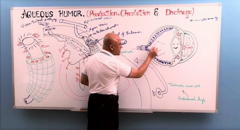
The ciliary processes produce aqueous humor in the posterior chamber. It flows through the pupil into the anterior chamber and drains through the trabecular meshwork into the canal of Schlemm and then the episcleral veins. Blocked drainage raises intraocular pressure and can cause glaucoma.
What happens in an oculomotor nerve palsy?
The eye rests “down and out” because the lateral rectus and superior oblique act unopposed. The upper eyelid droops (ptosis), and the pupil is dilated when the parasympathetic fibres are involved.
Which cranial nerve has the longest intracranial course?
The trochlear nerve (cranial nerve IV) has the longest intracranial course. It is also the thinnest cranial nerve and the only one that leaves the brainstem from its dorsal surface. It supplies the superior oblique muscle.
What happens in an abducens nerve palsy?
The abducens nerve supplies the lateral rectus, so the affected eye cannot move outward. The eye drifts inward and the patient sees double side by side, worst when looking toward the affected side.
How does the lacrimal gland get its nerve supply?
Parasympathetic fibres travel in the greater petrosal nerve to the pterygopalatine ganglion, then through the zygomatic nerve to the lacrimal nerve, which carries them to the gland. Sympathetic fibres arrive through the deep petrosal nerve.
What is the path of the visual pathway?
Light is detected in the retina, and signals travel through the optic nerve, optic chiasm and optic tract to the lateral geniculate nucleus. From there the optic radiation carries them to the primary visual cortex in the occipital lobe.
Questions about this course
Is the Dr. Najeeb eye anatomy and physiology course suitable for beginners?
Yes. Each lecture starts from the basics and builds step by step with hand-drawn diagrams, so no prior medical knowledge is needed. Students who already know the basics can use the course to revise the retina, the uveal tract, the eye nerves and the visual pathway.
How many lectures are in the Eye Anatomy and Physiology course?
The course has 36 lecture parts, from the structure of the eyeball to the five-part series on the visual system.
What topics does the eye anatomy and physiology course cover?
It covers the structure of the eyeball, the sclera and cornea, the uveal tract (choroid, ciliary body and iris), the retina, aqueous humor production and drainage, the blood supply of the eye, the orbital veins, the nerves that supply the eye and lacrimal gland, horizontal conjugate eye movements and the visual system.
Can I watch a free sample before I sign up?
Yes. A free “Try Us Free” account gives you access to sample lectures, so you can see the teaching style before you decide on a plan.
Are notes available for the eye lectures?
Handwritten notes are available for every lecture in this course. They are sold separately through the Notes add-on or the Combo Plan.
Do I get lifetime access?
Yes. The premium plan gives lifetime access to the lectures, including future lectures added to the library.
Can I download the lectures for offline viewing?
Offline viewing is available only through the official Dr. Najeeb Lectures app. The lectures cannot be downloaded as files.
Is this course useful for USMLE, PLAB, COMLEX or NEET PG?
Yes, for the eye anatomy, physiology and clinical correlates that these exams test. The course does not include a question bank or a certificate.
What is the refund policy?
Dr. Najeeb Lectures offers a 7-day refund. To request one, email support@drnajeeblectures.com.