
Gross Anatomy Head and Neck
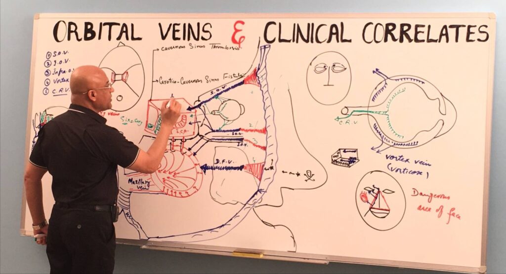
Dr. Najeeb’s Gross Anatomy Head and Neck course is a 78-part video lecture series on the cranial nerves, cavernous sinus, eye, ear, meninges and cervical lymph nodes. Every lecture is a hand-drawn, step-by-step whiteboard lesson by Dr. Najeeb, who has over 40 years of teaching experience. It is built for medical, dental, nursing and physiotherapy students who want head and neck anatomy explained through function and clinical cases instead of rote memorization.
| Course | Gross Anatomy Head and Neck |
|---|---|
| Instructor | Dr. Najeeb, over 40 years of teaching experience |
| Content | 78 lecture parts across eight modules |
| Teaching style | Hand-drawn, step-by-step whiteboard lectures |
| Level | Beginner-friendly; no prior medical knowledge needed |
| Access | Lifetime access with the premium plan, including future lectures |
| Notes | Handwritten notes for almost every lecture, sold separately (Notes add-on or Combo Plan) |
| Free preview | Available through a free “Try Us Free” account |
| Suitable for | Medical, dental, nursing, pharmacy and physiotherapy students; USMLE Step 1, PLAB, COMLEX and NEET PG preparation |
What you will learn in the head and neck anatomy course
The course follows the way head and neck anatomy is tested and used in clinical practice: each nerve, ganglion and structure is taught with its course, its connections and what happens when it is damaged.
- Cranial nerves III to X: course, branches, functions and lesions
- The trigeminal system and the ophthalmic and maxillary nerves
- The facial nerve and its clinical correlates, including facial palsy
- Head ganglia and autonomic pathways: ciliary, pterygopalatine and otic ganglia
- The cavernous sinus, its contents and cavernous sinus syndrome
- The structure and blood supply of the eye, including the sclera, cornea and uveal tract
- The middle ear, the auditory and vestibular systems and the vestibulo-ocular reflex
- The cervical lymph nodes and their seven levels
Clinical topics covered
The lectures keep returning to the clinical picture, so you learn the anatomy together with the findings you will meet in patients.
- Oculomotor, trochlear and abducens nerve palsies
- Cavernous sinus syndrome and cavernous sinus thrombosis
- Horner’s syndrome and the oculosympathetic pathway
- Facial nerve lesions and facial palsy
- Trigeminal pathways and sensory loss of the face
- Lacrimal gland innervation and the petrosal nerves
- Orbital veins and the spread of infection to the cavernous sinus
- Cervical lymph node levels used in neck examination and staging
Course curriculum: 78 head and neck anatomy lectures
| Module | Topics (number of lecture parts) | Parts |
|---|---|---|
| 1. Cranial nerves: basic concepts | Cranial Nerves: Basic Concepts (3) | 3 |
| 2. Cranial nerves III to X | Oculomotor Nerve and Clinical Correlates (6); Trochlear Nerve (2); Abducens Nerve (2); Introduction to the Trigeminal System (1); Trigeminal System (3); Ophthalmic Nerve: Lacrimal, Frontal and Nasociliary Nerves (1); Maxillary Nerve and its Branches (1); Facial Nerves (2); Facial Nerve and Clinical Correlates (4); Glossopharyngeal and Vagus Nerves (1) | 23 |
| 3. Head ganglia and autonomic pathways | Ciliary Ganglion and Connections (2); Pterygopalatine Fossa (1); Pterygopalatine Ganglion (2); Otic Ganglion (1); Petrosal Nerves: Greater, Deep and Lesser (1); Nerve Supply to the Lacrimal Gland (2); Oculosympathetic Pathway and Horner’s Syndrome (3) | 12 |
| 4. Cavernous sinus, meninges and orbital veins | Cavernous Sinus and Cavernous Sinus Syndrome (6); Meninges (2); Orbital Veins and Clinical Correlates (1) | 9 |
| 5. Eye structure and blood supply | Structure of the Eye Ball (1); Sclera (2); Cornea (1); Uveal Tract (3); Blood Supply to the Eye Ball (1); Ophthalmic Artery: Origin, Course and Branches (1) | 9 |
| 6. Middle ear | Anatomy of the Middle Ear (9) | 9 |
| 7. Inner ear, hearing, balance and eye movements | Auditory System (3); Vestibular System (3); Vestibulo-Ocular Reflex (2); Horizontal Conjugate Eye Movements (3) | 11 |
| 8. Cervical lymph nodes | Cervical Lymph Groups (1); Seven Levels of Cervical Lymph Nodes (1) | 2 |
Total: 78 lecture parts.
Who this course is for
- Medical students (MBBS, MD) learning head and neck anatomy for the first time
- Dental students who need the cranial nerves and regional anatomy of the head and neck
- Nursing, pharmacy and physiotherapy students
- Candidates preparing for USMLE Step 1, PLAB, COMLEX or NEET PG
No prior medical knowledge is needed. Each topic begins with the basic idea and builds up with diagrams.
How to study with this course
- Start with the three lectures on the basic concepts of cranial nerves, then move through nerves III to X in order.
- Watch each lecture with a blank page and redraw the diagram as Dr. Najeeb draws it.
- Use the handwritten notes for revision after you finish each module.
- Connect each nerve to its clinical lesion, as the lectures do, to remember it for exams.
Pair this course with the Neuro Anatomy & Neuro Physiology course for the brain and spinal cord, the Eye course for ophthalmology, Embryology for the development of the head and neck, and Histology for the tissues involved. To go further, add Clinical Medicine for trigeminal neuralgia, Horner syndrome and cervical lymph node levels, Upper Limb for the brachial plexus at the root of the neck, Nervous System Pathology for meningitis and neuromuscular disease, Respiratory Pathology for the Pancoast tumor and Microbiology for the infections of the head and neck.
Head and neck anatomy in brief
Which cranial nerve has the longest intracranial course?
The trochlear nerve (cranial nerve IV) has the longest intracranial course. It is also the thinnest cranial nerve and the only one that leaves the brainstem from its dorsal surface. It supplies the superior oblique muscle of the eye.
What does the trochlear nerve do?
The trochlear nerve supplies the superior oblique muscle, which rotates the eye inward (intorsion) and helps move it downward, especially when the eye is turned inward. A lesion causes vertical double vision that is worst when looking down and in, such as when reading or walking downstairs.
What happens in an oculomotor nerve palsy?
The eye rests “down and out” because the lateral rectus and superior oblique act unopposed. The upper eyelid droops (ptosis), and the pupil is dilated when the parasympathetic fibres are involved.
What passes through the cavernous sinus, and what is cavernous sinus syndrome?
The internal carotid artery and the abducens nerve pass through the sinus. The oculomotor, trochlear, ophthalmic (V1) and maxillary (V2) nerves run in its lateral wall. Cavernous sinus syndrome combines eye-movement paralysis with double vision, numbness over the upper face and often eye bulging (proptosis), usually from thrombosis, infection or a tumour.
What are the functions of the facial nerve?
The facial nerve supplies the muscles of facial expression and the stapedius, carries taste from the front two thirds of the tongue through the chorda tympani, and gives parasympathetic supply to the lacrimal, submandibular and sublingual glands.
What is Horner’s syndrome?
Horner’s syndrome is a triad of ptosis, a constricted pupil (miosis) and reduced sweating (anhidrosis) on one side of the face. It results from interruption anywhere along the three-neuron oculosympathetic pathway from the hypothalamus to the eye.
What are the seven levels of cervical lymph nodes?
Level I: submental and submandibular. Level II: upper jugular. Level III: middle jugular. Level IV: lower jugular. Level V: posterior triangle. Level VI: central compartment. Level VII: superior mediastinum.
How does the lacrimal gland get its nerve supply?
Parasympathetic fibres travel in the greater petrosal nerve to the pterygopalatine ganglion, then through the zygomatic nerve to the lacrimal nerve, which carries them to the gland. Sympathetic fibres arrive through the deep petrosal nerve.
Questions about this course
Is the Dr. Najeeb head and neck anatomy course suitable for beginners?
Yes. Each lecture starts from the basics and builds step by step with hand-drawn diagrams, so no prior medical knowledge is needed. Students who already know the basics can use the course to revise cranial nerves, the eye, the ear and clinical correlates.
How many lectures are in the Gross Anatomy Head and Neck course?
The course has 78 lecture parts, from the basic concepts of cranial nerves to the seven levels of cervical lymph nodes.
What topics does the head and neck anatomy course cover?
It covers cranial nerves III to X, the head ganglia and autonomic pathways, the cavernous sinus and cavernous sinus syndrome, the meninges, the structure and blood supply of the eye, the middle ear, the inner ear (hearing and balance), eye movements, Horner’s syndrome and the cervical lymph nodes.
Can I watch a free sample before I sign up?
Yes. A free “Try Us Free” account gives you access to sample lectures, so you can see the teaching style before you decide on a plan.
Are notes available for the head and neck lectures?
Handwritten notes are available for almost every lecture in this course. They are sold separately through the Notes add-on or the Combo Plan.
Do I get lifetime access?
Yes. The premium plan gives lifetime access to the lectures, including future lectures added to the library.
Can I download the lectures for offline viewing?
Offline viewing is available only through the official Dr. Najeeb Lectures app. The lectures cannot be downloaded as files.
Is this course useful for USMLE, PLAB, COMLEX or NEET PG?
Yes, for the head and neck anatomy and clinical correlates that these exams test. The course does not include a question bank or a certificate.
What is the refund policy?
Dr. Najeeb Lectures offers a 7-day refund. To request one, email support@drnajeeblectures.com.